
Ovalbumin(OVA) induced asthma model
Animal Model - Asthma Model
From ELK Biotech Wechat public account
Ovalbumin (OVA) induced asthma model

Background
Asthma (Asthma) is a chronic airway inflammatory disease characterized by variable airflow obstruction, airway hyperreactivity (AHR), and airway inflammation. The main clinical signs of asthma are shortness of breath, wheezing, coughing, and increased mucus production after contact with allergens. Its onset is caused by a complex interaction between genetic, epigenetic and environmental factors.
Different pathological changes are mediated by a variety of cells involved in immune response, such as airway epithelial cells, eosinophils and T lymphocyte subsets. Among them, Th2 cells are considered to be dominant in high eosinophilic asthma, accompanied by increased levels of cytokines IL-4, IL-5 and IL-13. The classic asthma model is the ovalbumin (OVA)-induced airway inflammation model, which is sensitized by multiple intraperitoneal injections of OVA and stimulated by aerosol inhalation of OVA to establish an acute asthma model.
Experimental materials and methods
01
Experimental material
·SD rat(Male, 6-8 weeks)
·Ultrasonic nebulizer(Manufacturer: Fulin Item No.: W001)
·Ovalbumin(Manufacturer: McLean Item Number: E6377)
·aluminum hydroxide(Manufacturer: Shanghai test Chinese medicine code: 20001060)
02
Modeling method
On the 1st and 8th day, normal saline containing 10 mg ovalbumin and 200 mg aluminum hydroxide were injected intraperitoneally. On the 15th day, normal saline containing 1% ovalbumin was inhaled for 30 minutes once a day for 14 days.
03
Evaluation index
Serum inflammatory factors and IgE were detected and HE staining was performed.
Verification
Detection of serum inflammatory factors and IgE

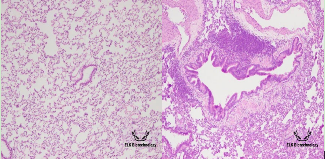
HE staining

Normal group Model group
HE Staining Results:In the normal group, the alveolar structure was normal and the bronchial epithelial structure was basically intact. In the model group, the alveolar wall was obviously thickened, the alveolar space was narrowed, the interval was widened, and the structure was disordered. A large number of inflammatory cells were seen in the bronchi, perivascular, and pulmonary interstitium. Bronchial smooth muscle and basal lamina thickened obviously, goblet cells proliferated and airway mucosa proliferated irregularly.


