


COPD Model-Experiment Sharing
 source:ELK Biotechnology
 date:2024-12-30
 views:1789
1.Background
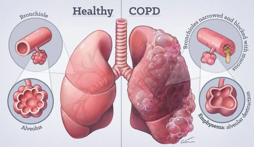
Model of chronic obstructive pulmonary disease Chronic obstructive pulmonary disease (COPD) is a common respiratory system disease with high incidence of a disease and mortality, which may lead to progressive pulmonary dysfunction, respiratory failure and cor pulmonale.
The cause of COPD is related to abnormal inflammatory response to harmful gases and particles, such as smoking, occupational exposure to dust and chemicals, air pollution, etc. Determination of the cause is particularly important for the prevention and treatment of COPD.
In order to study the pathogenesis of COPD and find effective treatment, the establishment of dynamic object model is an effective tool for scientific research.
2.Experimental Materials & Methods
Experimental material: C57 mice (male, 6-8 weeks); Cigarettes (Manufacturer: Honghe)
Modeling method:
Adaptive feeding for 7 days, the mice were placed in a self-made smoking poison box by fumigation. The top of the box had a small hole of 1.5X1.5cm to keep the air inside the box connected with the outside.
Lighting Honghe cigarettes (tar 11mg, nicotine 1.0mg, carbon monoxide 13mg), Put the mice into the box, close the box, take out the mice after 1 hour, rest for 15 minutes, light the cigarette again, continue to put the mice in the box for 1 hour, smoke once a day, smoke for 2 hours each time, smoke for 6 days every week, model 20 weeks after the material, do HE staining.
Evaluation indicators: Alveolar lavage fluid inflammatory factor detection, HE staining.
3.Verification
◆ HE staining
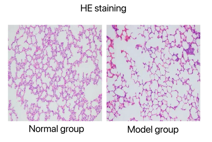
HE staining results: The structure of alveolar wall of lung tissue in normal group was continuous and relatively complete. In the model group, the bronchial wall and surrounding inflammatory cells were infiltrated, epithelial cells fell off, and showed villous hyperplasia. Pulmonary interstitial and alveolar cavity inflammatory cell infiltration, individual visual field bleeding, pulmonary interstitial proliferation; The normal structure of the alveoli was destroyed, part of the alveoli were broken and fused with each other to form a larger alveolar cavity.
◆ Detection of inflammatory factors in bronchoalveolar lavage fluid
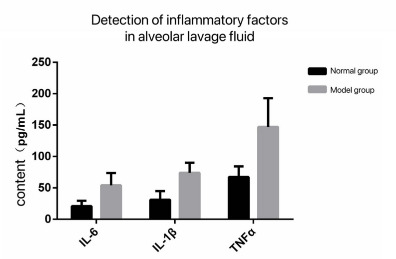
Results of serum inflammatory factor detection: The levels of serum IL6, IL-1β, TNFa and other inflammatory factors in the model group were significantly increased compared with those in the normal group.

 RETURN