


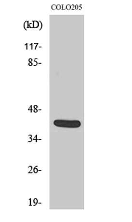
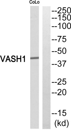


Vasohibin rabbit pAb
 One-click to copy product information
 Size: Size: |
 Price: Price: |
|---|---|
| 50 µL | $148.00 |
| 100 µL | $248.00 |
One-click to copy product information| Size: |
Price: |
|---|---|
| 50 µL | $148.00 |
| 100 µL | $248.00 |
 Manual