
Bleomycin-induced pulmonary fibrosis model
1.Research Background
Pulmonary fibrosis is a chronic, progressive lung disease characterized by the proliferation of fibroblasts and excessive accumulation of extracellular matrix, accompanied by inflammatory damage and structural destruction of lung tissue. This condition represents the end-stage changes of a range of pulmonary diseases, leading to abnormal repair of lung tissue, destruction of alveolar structures, and fibrosis. Pulmonary fibrosis severely impacts respiratory function, manifesting as dry cough and progressive shortness of breath. As the disease and lung damage progress, respiratory function deteriorates further. Idiopathic pulmonary fibrosis (IPF) is increasingly prevalent and has a higher mortality rate compared to most cancers, often referred to as a "cancer-like disease." Since the majority of pulmonary fibrosis cases have unknown causes, it is crucial to establish animal models that closely resemble the pathological process of human pulmonary fibrosis to study its mechanisms, pathological processes, and drug efficacy.
2.Experimental Materials and Methods
Experimental Materials: SD rats (male, 180-220g); Bleomycin (MCE, HY-17565A).
Modeling Method: 1. Adapt animals by feeding them for 7 days. Anesthetize with pure oxygen using an anesthesia machine. Shave the neck hair of the rats and disinfect with iodine tincture. 2. Expose the trachea and inject 5 mg/kg of bleomycin solution into the trachea. After injection, immediately rotate the rat upright for 30 seconds to ensure even distribution of the solution in the lungs, then gently place the rat back in the cage to lie flat and wake up naturally. Maintain normal diet and collect samples 4-5 weeks post-modeling for HE staining and Masson staining.
Evaluation Indicators: Biochemical analysis of lung tissue, Masson staining, HE staining.
3.Validation
◆ Masson Staining

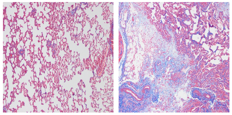
Normal Group Model Group
Masson staining results show that, compared to the normal group, the model group exhibits extensive collagen fiber proliferation in lung tissue, which stains blue. The alveolar walls are markedly thickened, and there is significant destruction of alveolar structures, with alveolar cavities reduced and becoming more solid. Fibrosis is observed in both alveoli and interstitium, with collagen deposition.
◆HE Staining

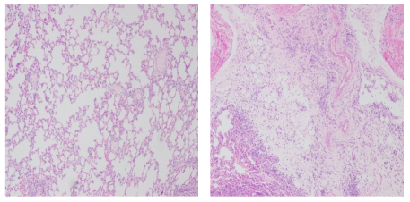
Normal Group Model Group
HE staining results reveal that, compared to the normal group, the model group has disorganized lung tissue structure, with destruction of alveolar structures and alveolar cavities reduced or absent. There is substantial infiltration of inflammatory cells in the alveolar septa and cavities. The alveolar walls are thickened, and the septa are widened.
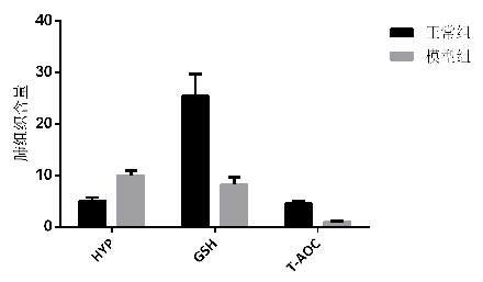
◆ Biochemical Analysis of Lung Tissue


