
Animal modeling-pancreatic cancer liver metastasis model Background introduction
The comprehensive treatment of pancreatic cancer, mainly surgery, is not effective, and metastasis occurs early after surgery. The main cause of death from pancreatic cancer is metastatic disease caused by pancreatic cancer, and the most common site is the liver.
Establishing a good animal model of liver cancer has a good alternative role in simulating the formation of human liver cancer and anti-tumor immune response research. Currently, there are many models such as subcutaneous transplanted tumors, orthotopic transplanted tumors, and pancreatic cancer liver metastasis tumors for scientific research applications.
Experimental materials & methods
Experimental materials
C57 mice (male, 6-8 weeks), PancO2 cells (transfected with luciferase)
Modeling method
Prepare a cell suspension.
After anesthetizing C57 mice, disinfect with 0.5% iodine.
Slowly inject the cell suspension into the middle of the lower pole of the spleen, and the spleen capsule can be seen to swell and turn white rapidly.
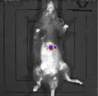
The tumor successfully metastasized to the liver and formed obvious lesions (time is 3 weeks-4 weeks), and the pancreatic and liver tumor formation status was observed based on in vivo imaging.
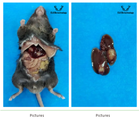
At the end of the cycle, liver tissue was promptly removed and HE staining was used to detect liver tissue pathology.
Evaluation indicators
In vivo imaging, gross observation, HE staining
Verification

In vivo imaging
Gross observation

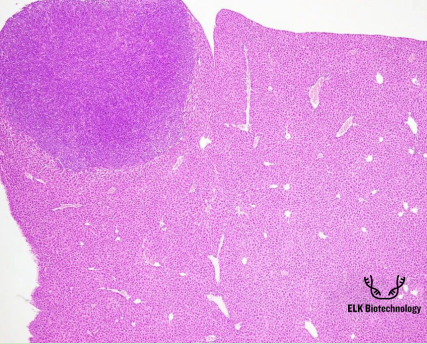
HE staining

Pictures
HE staining results: The liver tumor is nodular in shape, with abnormal proliferation forming a local mass, which is clearly demarcated from the surrounding tissue. Tumor cells are distributed in clusters with obvious heterogeneity. The nuclei of tumor cells in the tumor area are large, numerous, and deeply stained. Nuclear division and pyknosis can be seen in some fields of view, and the nucleoli are enlarged and increased in number.


