


Tumor necrosis factor-a,TNF-a
 source:ELK Biotechnology
 date:2023-08-09
 views:4779
Definition
Definition 1: Mainly produced by activated monocytes/macrophages, can kill and inhibit tumor cells, promote neutrophil phagocytosis, resist infection, cause fever, induce hepatocyte acute phase protein synthesis, and promote myeloid leukemia cells to macrophages, Phage differentiation, promoting cell proliferation and differentiation, is an important inflammatory factor and is involved in the pathological damage of some autoimmune diseases.
Definition 2: A cytokine naturally produced by macrophages in response to bacterial infection or other immune sources. It works synergistically with interferon to kill tumor cells.
Definition3: Cytokines that are mainly produced by activated monocytes/macrophages which can kill and inhibit tumor cells. Promotes phagocytosis of neutrophils, resists infection, causes fever, induces acute phase protein synthesis in hepatocytes, promotes the differentiation of myeloid leukemia cells into macrophages, promotes cell proliferation and differentiation, is an important inflammatory mediator, and is involved in certain autoimmunity Pathological damage of disease.
Tumor necrosis is a small protein secreted by macrophages. TNF-a is mainly secreted by monocytes-macrophages; TNF-b is mainly secreted by activated T lymphocytes, and the two have similar pyrogenicity. Small doses showed unimodal fever, and large doses showed bimodal fever; TNF could stimulate the production of IL-1 both in vitro and in vivo. Not heat resistant, inactivated at 70℃ for 30min.
Introduction
In 1975,Carswell et al. found that after injection of LPS in mice inoculated with BCG, the serum contained a factor that can kill certain tumor cells or cause blood necrosis of tumor tissue in vivo, called tumor necrosis factor(tumor necrosis factor, TNF). In 1985,Shalaby named the TNF produced by macrophages as TNF-a and the lymphotoxin(LT) produced by T lymphocytes as TNF-b. TNF-a is also known as cachexia.
Production of TNF

TNF-a is a monoline, mainly produced by monocytes and macrophages, and LPS is a strong stimulator,IFN-IFN-γ、M-CSF、GM-CSF can stimulate the production of TNF-a by monocytes/macrophages, while PGE can inhibit them. Promonocytic cell line U937 and promyeloid cell line HL-60 can produce higher levels of TNF-a when stimulated by PMA. T lymphocytes, T cell hybridomas, T lymphoid cell lines and NK cells also can secrete TNF-a under the stimulation of PMA,SAC,PMA and anti-IgM can stimulated normal B cells to produce TNF-a. In addition, neutrophils, LAK, stellate cells, endothelial cells, smooth muscle cells can also produce TNF-a.
TNF-b is a lymphokine, both of antigens and mitogens can stimulate T lymphocytes to secrete TNF-b. PMA stimulated RPMI1788B lymphoblasts to secrete high levels of TNF-b.
Molecular structure and gene of TNF

1)The human TNF-a gene is about 2.76kb in length, and the mouse is 2.78kb. The structure is very similar, Both of them consist of 4 exons and 3 introns, which are closely linked to the MHC gene group and are located in the first 6 and 17 pairs of chromosomes. In 1984, rHu TNF-a cDNA was successfully cloned from HL-60,U937 and other cells, and was highly expressed in E.coli. Human TNF-a precursor is composed of 233 amino acid residues, including a signal peptide of 76 amino acid residues, and the mature TNF-a after excision of the signal peptide is 157 amino acid residues, non-glycosylated, positions 69 and 101 two cysteines form an intramolecular disulfide bond. The molecular weight of rHu TNF-a is 17KDa. The precursor of mouse TNF-a is 235 amino acid residues, the signal peptide is 79 amino acid residues, and the molecular weight of mature mouse TNF-a (rMuTNF-a) is 17KDa, which consists of 156 amino acid residues, positions 69 and 100 two cysteines form an intramolecular disulfide bond and have a glycosylation point, but glycosylation does not affect its biological function, rHu TNF-a has 79% amino acid homology with rMu TNF-a, and the biological effect of TNF-a seems to have no obvious species specificity. Recently, it was reported that 155 amino acid human TNF-a with 2 amino acids(Val,Arg) less at the N-terminal was expressed by genetic engineering technology, which has better biological activity and anti-tumor effect. In addition, genetic engineering methods were used to delete 7 amino acid residues at the amino end of TNF-a molecule, and then 8Pro,9Ser and 10Asp were changed to 8Arg,9Lys and 10Arg,or 157 Leu was changed to 157Phe at the same time. The activity of TNF-a in killing L929 cells in vitro was about 1000tims higher than that of natural TNF. The effect of tumor hemorrhage and necrosis was also significantly increased in vivo. The natural forms in which TNF-a and b exert their biological effects are the homotrimers.
2)Human and mouse TNF-b genes are located on chromosomes 6 and 17 respectively. The HuTNF-b molecule consists of 205 amino acid residues and contains a signal peptide of 34 amino acid residues. The mature HuTNF-b molecule has 171 amino acid residues and a molecular weight of 25KDa. The rHu TNF-b molecule consists of 202 amino acid residues, including a signal peptide of 33 amino acid residues, and the mature molecule has 169 amino acid residues, which has 79% homology with HuTNF-b. Hu TNF-b and Hu TNF-a have 56% DNA homology and 36%homology at the amino acid level.
TNF receptors
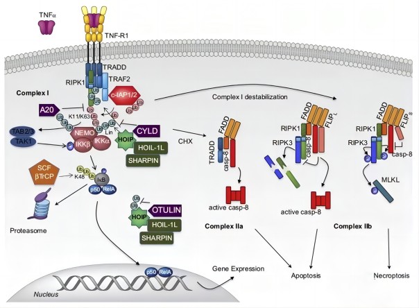
1)Types of TNF-R
TNF-R can be divided into two types: type I TNF-R,55KDa, CD120a, 439 amino acid residues, this type of receptor may play a major role in cytolytic activity: type II TNF-R, 75KDa,CD120b, 426 amino acid residues
based ,this type of receptor may be involved in signaling and T cell proliferation. Both types of TNF-R include three parts: the extracellular membrane region, the transmembrane region and the cytoplasmic region. The extracellular membrane region has 28%homology,but there is no homology in the cytoplasmic region, which may be different from the mediation of signal transduction pathways. TNF-R belongs to the nerve growth factor receptor(NGFR) superfamily. The receptors for TNF-a and TNF-b may be the same. TNF-R exists on the surface of a variety of normal and tumor cells. Generally, the number of receptors per cell is 103 to 104. For example, the ME-180 tumor cell line TNF-aR is about 2000per cell, and the Kd is 2*10-10M. The numbers and affinities of different cell surface TNF-aRs do not appear to parallel the sensitivity of cells to TNF-a. The mechanism of signal transmission after TNF-a binds to the corresponding receptor is not clear, it may be related to the activation of protein kinase C(PKC),which catalyzes the phosphorylation of receptor protein.
2)Soluble TNFR
TNF binding protein(TNF-BP) is the soluble form of TNFR, there are two kinds of sTNFRI (TNF-BPI) and sTNFR II(TNF-BP II). It’s generally believed that sTNFR has the effect of limiting TNF activity, or stabilizing TNF, and has an important regulatory role in the cytokine network. Seckiner in 1988 found that there are TNF inhibitors in the urine of febrile patients with a molecular weight of 33KDa. Olsson in 1989 also found TNF-BP in the blood and urine of patients with chronic renal insufficiency. TNF-BP can specifically bind to TNF and inhibit the activity of TNF, such as inhibiting its cytotoxic activity and inducing the production of IL-1, which can promote the growth of subcutaneously inoculated Meth A botulinum, which can be found in normal pregnancy urine. Elevated in inflammation, endotoxemia, meningococcal infection, SLE,HIV infection, renal insufficiency and tumors. Soluble TNFR can effectively alleviate the pathological changes of adjuvant arthritis and septic shock.
Biological activity of TNF
The biological effects of TNF-a and TNF-b are very similar, which may be related to the similarity of molecular structure and the identity of receptors. But there are differences in certain biological roles.
1)Kill or inhibit tumor cells
TNF can kill some tumor cells in vivo and in vitro(cytolytic action),or inhibit proliferation(cytostatic action). The sensitivity of tumor cell lines to TNF-a varies greatly, and the TNF-a even has stimulating effect on very few tumor cells. Treating tumor cells(such as mouse fibroblast line L9290 with actinomycin D, mitomycin C, cycloheximide, etc. Can significantly enhance the activity of TNF-a in killing tumor cells. Tumor response to TNF-a in vivo also varied widely, not in parallel with the sensitivity of their in vitro cell lines to TNF-a. The same cell line may have susceptible and resistant strains such as L929-S and L929-R. In addition, the expression of endogenous TNF in target cells may make cells resistant to the cytotoxic effects of exogenous TNF. Therefore, the sensitivity of cells to exogenous TNF can be altered by inducing or inhibiting the expression of endogenous TNF. Macrophage membrane-bound TNF may be involved in the killing of target cells.
The mechanism of TNF killing tumor is not very clear. Compared with complement or perforin(perforin) killing cells, TNF killing cells have no perforation phenomenon, and the killing process is relatively slow. The killing of tumor tissue cells by TNF may be related to the following mechanisms.
a.direct killing or inhibition
After TNF binds to the corresponding receptor, it moves into the cell and is taken up by the lysosome of the target cell, resulting in a decrease in the stability of the lysosome and the leakage of various enzymes, causing cell lysis. It’s also believed that TNFN activates phospholipase A2,releases superoxide and causes DNA fragmentation. And the phospholipase A2 inhibitors can reduced the anti-disease effect of TNF. TNF can change target cell glucose metabolism, reduce intracellular pH, and lead to cell death.
b.Through the regulating effect of TNF on the immune function of the body, promote the killing of tumor cells by T cells and other killer cells.
c.TNF acts on vascular endothelial cells, damages endothelial cells or leads to vascular dysfunction, vascular damages and thrombosis, resulting in local blood flow blockage in tumor tissue, resulting in hemorrhage and hypoxia necrosis.
2) Improve the phagocytic ability of neutrophils
Increases superoxide anion production, enhances ADCC function, stimulates cellular degranulation and secretion of myeloperoxidase. TNF pre-cultured with endothelial cells can increase the expression of MHC I antigens, ICAM-1, the secretion of IL-1.GM-CSF and IL-8. And promote the adhesion of neutrophils to endothelial cells, thereby stimulating the body in local inflammatory response, the induction effect of TNF-a is stronger than that of TNF-b. TNF stimulated monocytes and macrophages to secrete IL-1, and regulates the expression of NHC II antigens.
3)Anti-infection
For example, it inhibits the growth of Plasmodium, inhibits virus replication(such as adenovirus type II, herpes virus type II), inhibits viral protein synthesis, virus particle production and infectivity, and can kill virus-infected cells. The antiviral mechanism of TNF is not very clear.
4)TNF is an endogenous pyrogen
Causes fever and induces the synthesis of acute phase proteins in hepatocytes. TNF induced fever may be caused by directly stimulating the hypothalamic thermoregulatory center and stimulating the release of IL-1 form macrophages. It can also stimulate other cells to produce IL-6 through IL-1 and TNF-a.
5)Promote the differentiation of myeloid leukemia cells into macrophages
Such as promoting the differentiation of myeloid leukemia cells ML-1,monocytic leukemia cells U937, and promyelocytic leukemia cells HL60,the mechanism is unclear. TGF-b can inhibit various biological activities of TNF-a, but does not inhibit the induction of TNF-a on the differentiation of myeloid leukemia cells, and even has synergistic effect.
6) Promote cell proliferation and differentiation
TNF promotes the expression of NHC I antigens in T cells, enhances IL-2 dependent thymocytes and T cell proliferation, promotes the production of lymphokines such as IL-2,CSF and IFN-γ, and enhances mitogens or foreign antigens to stimulate B cell proliferation growth and Ig secretion. TNF-a has a growth factor-like effect on some tumor cells, and synergizes with the pro-proliferative effects of EGF,PDGF and insulin, and promotes the expression of EGF receptors. TNF can also promote the expression of proto-oncogenes such as c-myc and c-fos, which are closely related to cell proliferation, and cause the transition of the cell cycle from G0 phase to G1 phase. Recently, it has been reported that INF-b(LT) is an autocrine growth factor of EB virus-transformed lymphoblasts. Anti-LT antibodies, sTNfR and TNF-a can inhibit the proliferation of EB virus-transformed lymphocytes.
IL-1, IFN-γ and GM-CSF can significantly enhance the biological effects of TNF, which may be related to the increased expression of cellular TNF receptors. An anti-TNF-a monoclonal antibody has been reported, which can mimic some of the biological effects of TNF-a, which has not been seen in other factors.
TNF and clinical
The application of TNF in the treatment of tumors has begun clinical phase II trials, and it can also be combined with IL-2 to treat tumors. Currently, it is believed that the efficacy of systemic drugs is not as good as that of local drugs. The latter, such as intralesional injection, has high local concentrations and less side effects. In recent years, TNF gene therapy has been used for clinical validation of melanoma and other tumors. It is worth nothing that TNF is related to the occurrence of some clinical diseases.
1)Septic shock: It is currently believed that disseminated intravascular coagulation and toxic shock caused by Gram-negative bacilli or meningococcus are caused by bacterial endotoxin stimulating the body to produce excessive TNF-a, causing fever and serious damage to the heart and adrenal glands, respiratory and circulatory failure, even cause death. The level of TNF is positively correlated with mortality. The pathogenesis may be that TNF stimulates endothelial cells, leading to inflammation, tissue damage and coagulation. TNF is also an important factor in acute liver necrosis. Peripheral blood cells in fulminant viral liver failure induce TNF, and the activity of IL-1 increase, which is related to the severity of the disease. The mechanism of TNF-mediated endotoxic shock is still unclear. It is believed that TNF can promote the production of prothrombin active substances and inhibit endothelial cell thrombin to regulate toxic shock. Antibodies to TNF (antisera or monoclonal antibodies) are effective in preventing lethal endotoxin in vivo in mice, rabbits and baboons. The application of anti-TNF McAb in the treatment of sepsis and septic shock has entered the phase III clinical trial, and the anti-TNF chimeric antibody in the treatment of bacterial infection has also started the phase I clinical trial.

2)Cachectin: TNF-a also known as cachetin which can induce cachexia in the body.

3)The relationship between TNF and viral replication: TNF also has antiviral effects similar to IFN, preventing the synthesis of early viral proteins, thereby inhibiting viral replication, and synergistically antiviral effects with TNF-a and xn--tnf--emd.On the other hand, TNF induces HIV-I gene expression in T cells. TNF and HIV infected CD4+ cells activate or induce NF-kB binds to the enhancer site of HIV’s long terminal repeat (LTR) and activates HIV genes, which may be related to the pathogenesis of AIDS. The production of TNF-a in monocytes in AIDS patients is increased, and the level of TNF-a in serum is increased.
Related products
TNFa
Human TNFa(Tumor Necrosis Factor Alpha) ELISA Kit
Mouse TNFa(Tumor Necrosis Factor Alpha) ELISA Kit
Rat TNFa(Tumor Necrosis Factor Alpha) ELISA Kit
TNF-α Rabbit pAb www.elkbiotech.cn/pro/12087
TNF-α Rabbit pAb www.elkbiotech.cn/pro/12345
TNFa receptor
TNF-R1 Rabbit pAb www.elkbiotech.cn/pro/15836
Mouse TNFRSF1A(Tumor Necrosis Factor Receptor Superfamily, Member 1A) ELISA Kit

 RETURN