


Mouse Renal Ischemia‑Reperfusion Injury Model
 source:ELK Biotechnology
 date:2026-08-06
 views:529

1. Background
The renal ischemia‑reperfusion injury model simulates the physiological process in which blood flow is restored to the kidney after an ischemic episode. It serves as a vital experimental tool in medical research for exploring the mechanisms underlying kidney injury and repair. This model is generally established by clamping the bilateral renal arteries of mice using micro‑vascular clips, allowing researchers to observe changes in renal function and kidney pathological features.
2. Experimental Materials and Methods
Experimental Materials
- C57 mice (male, 6‑8 weeks old)
- Isoflurane (RWD Life Science, Cat. No. R510‑22‑10)
- General‑purpose small‑animal anesthesia machine (RWD Life Science, Model R580S)
Model Establishment Protocol
- Mice undergo 7‑day acclimatized feeding, followed by anesthesia using a small‑animal anesthesia machine with pure oxygen mixed with isoflurane.
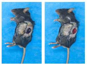
- A ventral skin incision is made to expose the kidneys. One renal pedicle is clearly visualized under direct vision, and the corresponding renal artery is dissected free. The respiratory status of the mouse is monitored throughout the ischemic phase, alongside renal color changes. The kidney turns bright red to dark red upon ischemia. After 45 minutes of ischemia, the ligature is released to initiate reperfusion. The abdominal wall is closed with layered sutures.
- Post‑operative animal wellbeing is monitored until full recovery from anesthesia. Kidney tissues are harvested at 24 hours after reperfusion.
Evaluation Indicators Macroscopic photographs of ischemic kidneys, serum biochemical assays, hematoxylin‑eosin (HE) staining, and terminal deoxynucleotidyl transferase‑mediated dUTP nick‑end labeling (TUNEL) fluorescence assay.
3. Model Validation
♦Macroscopic Observation of Renal Ischemia
♦HE Staining
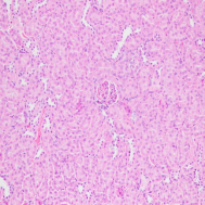
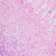
HE‑staining results: Compared with the normal group, the model group exhibits disorganized renal tissue architecture, with inflammatory cell infiltration in the renal interstitium. A large number of renal tubular epithelial cells show swelling, rupture and karyolysis. Widespread necrosis and exfoliation of tubular epithelial cells can be observed in severely damaged areas. Some renal tubules are dilated, and luminal casts formed by sloughed epithelial debris are present within the tubular lumen.
♦TUNEL Fluorescence Assay
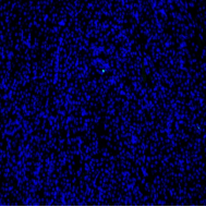
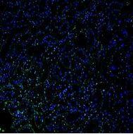
Note: Cell nuclei are stained blue by DAPI; TUNEL‑positive apoptotic cells exhibit green fluorescence. Compared with the normal group, massive apoptosis of renal tubular epithelial cells is detected in the model group.
♦Serum Biochemical Assays
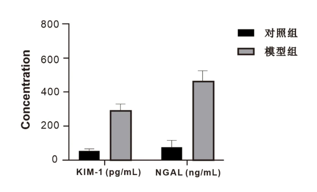
♦Serum ELISA Detection
| Catalog No. | Kit Name | Assay Target |
|---|---|---|
| BC020 | Creatinine (Cr) Assay Kit (sarcosine oxidase method) | Creatinine (Cr) |
| BC027 | Urea Assay Kit (urease method) | Urea |
| BC035 | Uric Acid (UA) Test Kit | Uric Acid (UA) |
| Catalog No. | Kit Full Name |
|---|---|
| ELK10195 | Mouse Neutrophil Gelatinase‑Associated Lipocalin (NGAL) ELISA Kit |
| ELK10659 | Rat Neutrophil Gelatinase‑Associated Lipocalin (NGAL) ELISA Kit |
| ELK038ES | EasyStep Human Neutrophil Gelatinase‑Associated Lipocalin (NGAL) ELISA Kit |
| ELK1063 | Human Neutrophil Gelatinase‑Associated Lipocalin (NGAL) ELISA Kit |
| ELK11098MS | Human Kidney Injury Molecule‑1 (KIM‑1) Microsample ELISA Kit |
| ELK11066MS | Mouse Kidney Injury Molecule‑1 (KIM‑1) Microsample ELISA Kit |
| ELK11066 | Mouse Kidney Injury Molecule‑1 (KIM‑1) ELISA Kit |
| ELK11098 | Human Kidney Injury Molecule‑1 (KIM‑1) ELISA Kit |
| ELK11099 | Rat Kidney Injury Molecule‑1 (KIM‑1) ELISA Kit |

 RETURN