


Collection, processing and storage of tissue homogenate and cell lysate samples
 source:ELK Biotechnology
 date:2024-12-30
 views:7370

Tissue homogenate
Tissue homogenization refers to an experimental technique in which tissue samples are broken up and evenly dispersed in buffer for subsequent experimental manipulation. In the following we will detail the procedure and precautions for tissue homogenization.
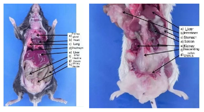
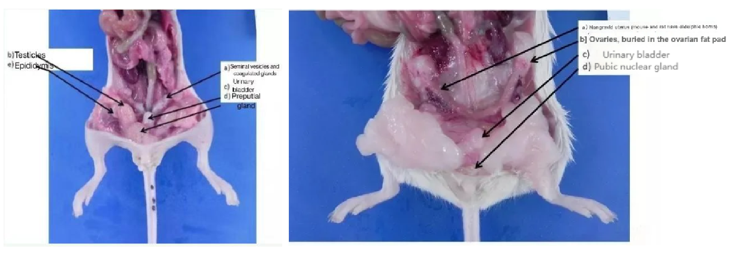
1. Take an appropriate amount of tissue and rinse with pre-cooled PBS (0.01mol/L, pH 7.0-7.4) to remove residual blood and impurities on the surface as much as possible. Specific mouse organs are shown below.
2.Weigh the tissue block (the mass to volume ratio of tissue to PBS is recommended to be 1:9, that is, 100mg of sample added to 900μl of PBS), and the tissue sampling volume is recommended to be more than 30mg at least to avoid too little homogenization buffer affecting the grinding effect.
3.Cut the weighed tissue into as small pieces as possible to fully homogenate; various homogenization methods can be used to achieve better crushing effect. Move the tissue block into the glass homogenizer and add the corresponding volume of PBS for full grinding (machine homogenate can be used in conditional laboratory). The process should be carried out on ice; the resulting homogenate can be further processed by ultrasonic crushing or repeated freezing and thawing (pay attention to ice bath cooling during ultrasonic crushing; repeated freezing and thawing method can be repeated twice).
4.The prepared homogenate was centrifuged at 5000×g for 5 minutes, and the supernatant was taken for detection. Since most of the tissue samples contain fat, if the supernatant was cloudy after homogenization, it could be stored at 4° appropriately. After the fat was solidified and stratified, the homogenate supernatant of the middle layer was taken for subsequent detection.
5.Tissue homogenates were determined for total protein content using a BCA protein quantification kit.(Since the material and grinding degree cannot be completely consistent, BCA quantification can be used as a correction means to make the subsequent results more accurate).
6.The 1:9 ground tissue homogenate samples generally need to be diluted for ELISA detection, and the optimal dilution ratio can be determined based on pre-experiments.
7.The final results of the ELISA experiments were divided by the corresponding BCA results to obtain the amount of target material per mg of total protein.
cell lysis buffer
Cell lysis is the process of destroying cell structure and releasing intracellular material, and in some experiments that need to detect intracellular proteins, cell lysate needs to be prepared for subsequent testing. The operated procedure for cell lysates was as follows:
1. Collection of cell precipitation:
① Suspension cells: For cells cultured in suspension, cell precipitation was collected directly by centrifugation, the culture medium was incubated at 1000 rpm at room temperature, centrifuged for 10 minutes, and the supernatant cells were discarded for precipitation.
② Adherent cells: For adherent cultured cells, digest the cells with trypsin, or scrape the cells with cells, put the culture medium at 1000 rpm at room temperature, centrifuge for 10 minutes, and discard the supernatant cells for precipitation.
Cell density should not be less than 106 cells / ml.
2. Washing of cell precipitation:
Add 0.5-1ml of PBS (isotonic) to the cell precipitate, centrifuged at 1000 RPM for 10 min, and discard the supernatant cells for precipitation. Repeat the above operation and wash it repeatedly for 1 to 2 times.
3. Add a certain amount of PBS to the cell precipitate (the amount of PBS is changed according to the measured indicators, it is recommended that 200-300ul) and add a small amount of mild lysate Triton X-100 (0.5%) to help lysis, lysis by sonication or repeated freeze-thaw.(Sonicated for about five minutes and centrifuged at 12000 x g for 5 minutes. Freeze and thaw repeatedly, freeze and thaw repeatedly with liquid nitrogen or -80℃ for 3-4 times, and then centrifuged at 12,000 xg for 5min). The supernatant was removed and assayed.
4. The lysate supernatant was used to determine the total protein content using the BCA protein Quantification kit. (Since the amount of cells and the degree of grinding could not be kept completely consistent, BCA quantification can be used as a correction to make subsequent results more accurate.)
5. The optimal dilution of ELISA was determined based on the pre-experiments.
6. The final results of the ELISA experiment divided by the corresponding BCA results can yield the amount of the target material per mg of total protein.
Note: Because many lysates are protein denaturants, which have a certain impact on the measurement of enzyme vitality, it is generally not recommended to use lysate for lysis, ELISA experiments with PBS lysis is enough.
Storage
After processing, the samples can be packed separately and sealed for storage. The storage should be less than 1 week at 4℃, no more than 1 month at -20 ℃, and no more than 3 months at -80℃. Be careful not to freeze and thaw repeatedly. The specimens should be slowly equilibrated to room temperature before use and should not be heated to melt.

 RETURN